
Reviewed by Dr. Amir Issa, MD, DABVLM, RPhS · CURA Vein Doctors · October 2026
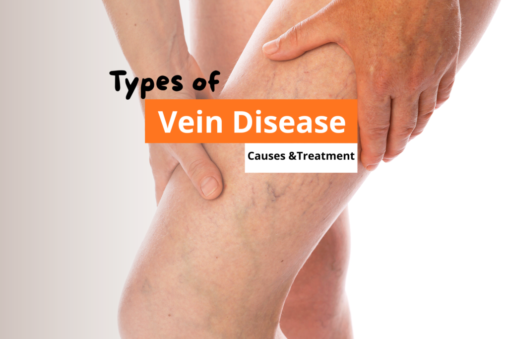
Most people discover they have a vein problem when they notice something on the surface: a blue cord behind the knee, a web of red lines near the ankle, unexpected puffiness by the end of the day. What they rarely know is that what they can see is usually the least of it. Vein disease in the legs spans a wide spectrum. At one end: cosmetic spider veins with no medical consequence. At the other: chronic venous insufficiency eroding the skin above the ankle, or a deep vein clot capable of reaching the lungs. Understanding which condition is present changes what treatment looks like and how urgently it is needed.
Vein Disease in Legs: What You’re Actually Dealing With
1. Varicose Veins
Valve failure in the saphenous system; bulging, twisted veins at CEAP C2
2. Spider Veins
Dilated capillaries at C1; cosmetic or feeder-linked; image before treating
3. Chronic Venous Insufficiency
Progressive venous hypertension; C2–C6; skin damage and ulcers at endpoint
4. Pelvic Congestion Syndrome
Internal varicosities; chronic pelvic pain; no visible leg findings
5. Superficial Thrombophlebitis
Clot + vein wall inflammation; 6–40% concurrent DVT; image above the knee
6. Deep Vein Thrombosis
Deep vein clot; pulmonary embolism risk; same-day evaluation required
1. Varicose Veins
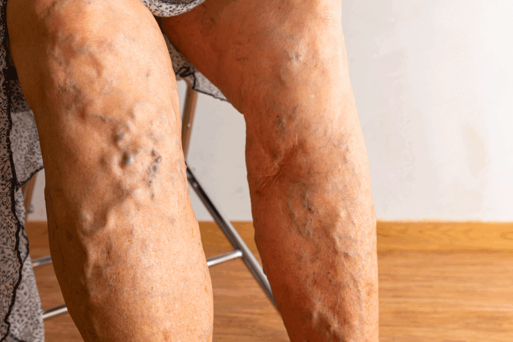
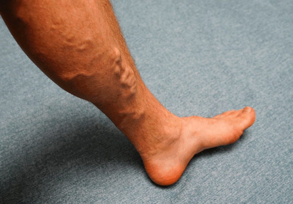
Varicose veins are enlarged, twisted veins that bulge visibly beneath the skin, most often at the back of the calf or along the inner thigh. They develop when valves inside the great or small saphenous vein stop closing properly. Blood that should move upward toward the heart falls back with each heartbeat. That pressure stretches the vein wall over time, producing the rope-like bulge that becomes visible at the surface.
They affect roughly 1 in 4 adults. The common assumption is that they are purely cosmetic. They are not. A bulging varicose vein is a visible sign that the venous system is under sustained, abnormal pressure. Left untreated, aching and heaviness worsen over years, and a significant portion progress to chronic venous insufficiency.
2. Spider Veins
Spider veins are smaller and sit much closer to the surface. They are dilated capillaries or venules, typically under 1 mm across, appearing as red, blue, or purple webs or fine lines on the thigh, calf, or ankle. They do not bulge. In the clinical CEAP classification they sit at C1, the lowest severity level in venous disease.
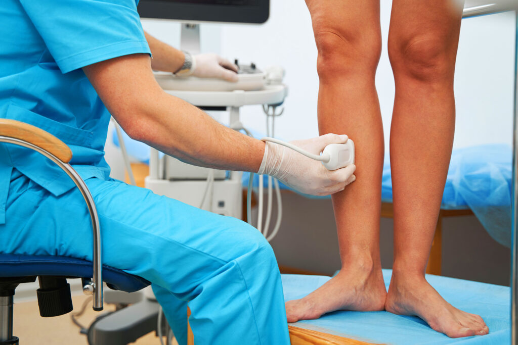
For many people they are cosmetic and never progress. For others, they mark the surface territory of a refluxing feeder vein lying beneath them. Treating spider veins with sclerotherapy without identifying and correcting that underlying source leads to rapid recurrence. This is why a duplex ultrasound before cosmetic vein treatment is more than a formality: it determines whether there is a deeper reflux point that needs to be addressed first, and whether what looks like a simple cosmetic issue has an underlying venous cause.
3. Chronic Venous Insufficiency
Chronic venous insufficiency (CVI) is the progressive disease that varicose veins, left unaddressed, often become. Sustained venous hypertension damages not just the veins but the tissue surrounding them. Fluid leaks into the lower leg. The ankle swells consistently rather than occasionally. The skin above the inner ankle begins to change: a brownish discoloration appears from hemosiderin deposited by red blood cells escaping through the capillary walls. This is frequently mistaken for aging, eczema, or a simple rash.
CEAP Severity Scale: How CVI Progresses
CVI is systematically underdiagnosed. In a 2020 international survey of 16,015 adults across eight countries, Rabe and colleagues found that only 28% of those with chronic venous disease had ever had a Doppler ultrasound. Most had been managing symptoms with compression or topical products for over a year before consulting a physician.

4. Pelvic Congestion Syndrome
Pelvic congestion syndrome is, in essence, varicose veins inside the abdomen. The ovarian and internal iliac veins dilate and reflux, producing chronic pelvic pain: a dull, persistent ache or pressure that worsens after prolonged standing, during menstruation, or after intercourse. Because it sits entirely inside the pelvis, it produces no visible surface abnormality on the legs and is easily attributed to gynecological causes for years.
Standard leg vein duplex ultrasound does not adequately image the pelvic veins. CT venography, MRI, or catheter-directed venography is needed for a confirmed diagnosis. The diagnostic path is longer and less familiar to most practitioners than the workup for leg vein problems. A vein specialist with pelvic venous training is the most direct route to diagnosis and treatment, which for most patients involves endovascular embolization of the affected pelvic veins.
5. Superficial Thrombophlebitis
Superficial thrombophlebitis is a clot inside a superficial vein combined with inflammation of the vein wall. The affected segment becomes tender, warm, red, and palpable as a firm cord under the skin. It most commonly occurs in varicose veins, where sluggish blood flow and damaged vein walls create conditions that favor clotting. It is painful and uncomfortable but typically resolves over days to weeks with anti-inflammatory medication and compression.
The clinical point that changes how it should be managed is the concurrent DVT rate. Studies report it at between 6% and 40%, depending on the anatomical location of the clot. A clot in the great saphenous vein close to the saphenofemoral junction carries the highest risk of extension into the deep venous system. For that reason, superficial thrombophlebitis presenting above the knee requires duplex ultrasound to rule out deep vein involvement. Reassurance without imaging is not appropriate when the clot is at that location.
6. Deep Vein Thrombosis
Deep vein thrombosis (DVT) is a blood clot in one of the deep leg veins, most commonly the popliteal, femoral, or iliac vein. It is the most serious condition in this group. The clot can detach and travel through the venous circulation to the lungs, causing a pulmonary embolism. Venous thromboembolism (DVT and PE combined) affects an estimated 900,000 Americans each year. Up to 100,000 die from it annually.
Leg signs can look identical to other venous conditions: unilateral swelling, calf pain, skin warmth. They can also be entirely absent. DVT is classified as provoked (triggered by surgery, immobility, cancer, or hormonal therapy) or unprovoked. Unprovoked DVT carries a higher recurrence risk and typically requires longer anticoagulation. A further complication: post-thrombotic syndrome, a form of CVI caused by valve destruction from the clot itself, develops in up to 50% of DVT patients. A person who has had one DVT is not simply back to their prior baseline after anticoagulation ends.
The three conditions most frequently confused at initial presentation are CVI, superficial thrombophlebitis, and DVT. All can present with a swollen, uncomfortable leg. The table below separates them by the features that matter most.
| Feature | Chronic venous insufficiency | Superficial thrombophlebitis | Deep vein thrombosis |
|---|---|---|---|
| How swelling presents | Bilateral; builds through the day; eases overnight | Localized to the affected vein segment; may not cause limb swelling | Typically unilateral; can be rapid in onset |
| Character of pain | Dull heaviness; worst by evening; present most days | Sharp, tender along a cord; worse with pressure or touch | Dull aching or absent; calf tenderness with compression |
| Skin changes | Brownish discoloration or thickening near the inner ankle | Red, warm streak directly over the affected vein | Redness or warmth may be present; no chronic skin changes early on |
| Duration pattern | Months to years; gradually progressive | Days to weeks; resolves with treatment | Acute onset; worsens without treatment |
| Key diagnostic test | Duplex ultrasound of the leg veins | Duplex ultrasound to exclude deep vein extension | Duplex ultrasound; urgent if PE symptoms are present |
| Urgency | Non-urgent; schedule evaluation within a few weeks | Moderate; image within 24 to 48 hours if above the knee | Urgent; same-day evaluation required |
When Vein Symptoms Have Gone Beyond Cosmetic
- Leg heaviness or aching that builds through the day and is present most days, not just during hot weather or after unusual activity
- Ankle swelling that does not fully clear after a night with your legs elevated
- Brownish skin discoloration, persistent itching, or a dry, leathery patch near the inner ankle
- Varicose veins that have appeared or visibly worsened in the past year
- A superficial vein that became tender, red, or hardened after a period of illness, travel, or reduced mobility
- Chronic pelvic pain in a woman with no confirmed gynecological cause, worse after prolonged standing
Any of these signals that the problem has a vascular component. Lifestyle measures will not stop the progression. A duplex ultrasound is the appropriate next step.
Vein Disease in Legs Symptoms: How They Build Over Time
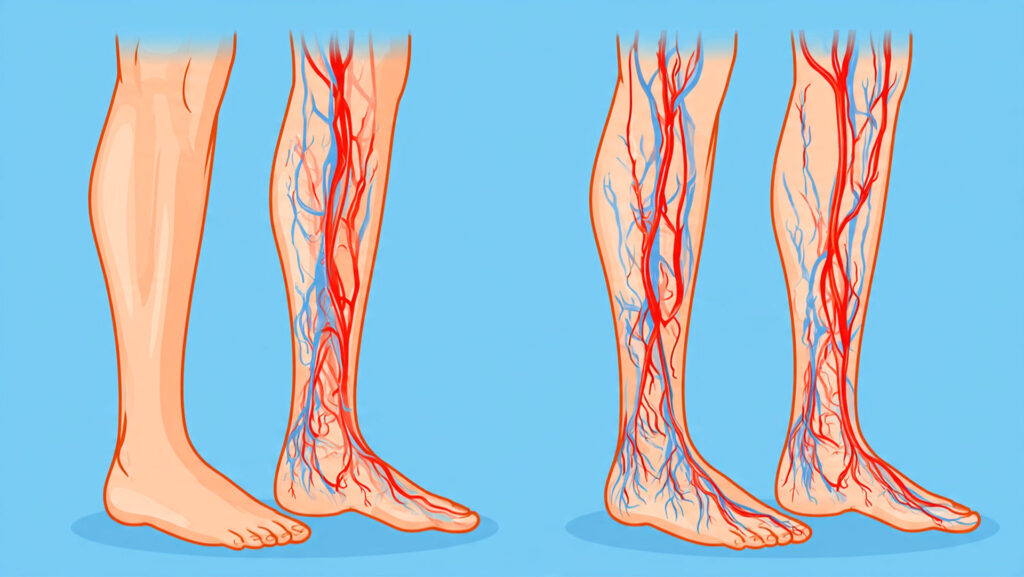
Venous symptoms rarely arrive suddenly. They build over months and years in a predictable sequence driven by rising venous pressure. Heaviness and aching come first, usually after prolonged standing or sitting. Mild ankle swelling follows, initially only in the evening, clearing overnight. The skin changes arrive later: brownish discoloration above the inner ankle, itching dismissed as dry skin, a patch of what looks like eczema that does not respond to treatment.
The problem with this slow progression is that each stage is easy to normalize. Tired legs at the end of a long day feel expected. Mild evening swelling in summer seems like heat. The skin patch becomes background noise. By the time a patient is at C4 with confirmed skin changes and measurable venous hypertension on duplex ultrasound, years have typically passed since the underlying reflux began. The window for preventing irreversible skin damage was open the whole time.
| What you are experiencing | Likely stage and approach | When to seek evaluation |
|---|---|---|
| Visible veins on the legs, no symptoms | C1 to C2; cosmetic or early reflux; duplex ultrasound before any cosmetic treatment | Elective; no urgency |
| Aching and heaviness that builds by evening, mostly better overnight | C2; active reflux likely; compression stockings and duplex evaluation | Within a few weeks |
| Ankle swelling that does not fully clear with legs elevated overnight | C3; ambulatory venous hypertension; compression plus imaging | Within 2 to 4 weeks |
| Brownish skin or persistent itching near the inner ankle | C4; prolonged venous hypertension with skin damage; imaging needed promptly | Within 1 to 2 weeks |
| Open wound or slowly healing sore near the ankle | C5 or C6; venous ulcer; specialist evaluation and wound management | Urgent; within days |
| Single warm, tender, red cord along a vein | Superficial thrombophlebitis; duplex ultrasound to exclude DVT extension | Within 24 to 48 hours if above the knee |
How to Treat Vein Disease in Legs
Treatment depends entirely on the type and stage. For varicose veins and CVI at stages C2 to C4, the first step is confirming reflux with duplex ultrasound, then eliminating the source. Endovenous laser ablation (EVLA) and radiofrequency ablation (RFA) close the refluxing vein segment from the inside using heat delivered through a thin catheter. Phlebectomy removes visible surface veins through small punctures. Sclerotherapy targets spider veins and residual smaller vessels. These are outpatient procedures with short recovery times and high long-term success rates when the reflux source is properly identified first.
DVT is treated with anticoagulation, the duration and agent depending on whether it is provoked or unprovoked and whether pulmonary embolism is involved. Superficial thrombophlebitis typically responds to compression, anti-inflammatory medication, and topical treatment, with imaging-guided reassessment if the clot is near the saphenofemoral junction. Pelvic congestion syndrome is treated with endovascular coil embolization or sclerotherapy of the affected pelvic veins, performed by an interventional radiologist or a vein specialist with pelvic venous training.
Seek Same-Day Evaluation For:
- Sudden swelling, warmth, or redness in one leg with calf tenderness (possible DVT)
- Any leg swelling combined with chest pain, shortness of breath, or a rapid heartbeat (possible pulmonary embolism)
- A cord-like vein near the groin becoming acutely tender or inflamed (risk of deep vein extension)
- An open wound or sore near the ankle that is not healing with standard wound care
- A leg that turns cold, pale, or mottled (possible arterial involvement)
Frequently Asked Questions
How to treat vein disease in legs?
Treatment depends on the type. Varicose veins and CVI are treated with endovenous ablation or sclerotherapy after duplex ultrasound confirms the reflux source. DVT requires anticoagulation; superficial thrombophlebitis responds to compression and anti-inflammatories; pelvic congestion syndrome to endovascular embolization.
How serious is chronic venous insufficiency?
At C2 to C3 it causes discomfort but is not life-threatening. At C4 and beyond it produces permanent skin changes and venous ulcers that recur unless the underlying reflux is corrected at its source.
Can you stop the progression of venous insufficiency?
Compression stockings, regular walking, and weight management significantly slow progression but do not reverse valve damage already done. Endovenous ablation changes the disease trajectory more fundamentally and is the appropriate intervention when reflux is confirmed on imaging.
What kind of exercise is recommended for venous insufficiency?
Walking is the most effective; it activates the calf muscle pump, the main driver of venous return from the legs. Swimming and cycling also engage it. Heavy weightlifting can worsen pelvic venous reflux and should be discussed with a vein specialist.
Recognize your symptoms in any stage above?
A duplex ultrasound identifies the source of reflux, maps the extent of valve failure, and rules out DVT in the deep system. CURA Vein Doctors offers vein assessments covered by most major insurance when venous symptoms are affecting daily function.
References
- Rabe E, Régnier C, Goron F, Salmat G, Pannier F. The prevalence, disease characteristics and treatment of chronic venous disease: an international web-based survey. J Comp Eff Res. 2020;9(17):1205–1218.
- Eberhardt RT, Raffetto JD. Chronic venous insufficiency. Circulation. 2014;130(4):333–346.
- Beckman MG, Hooper WC, Critchley SE, Ortel TL. Venous thromboembolism: a public health concern. Am J Prev Med. 2010;38(4 Suppl):S495–501.