

By CURA Vein Doctors · Last updated September 2026 · Compiled from peer-reviewed vascular medicine literature and clinical diagnostic guidelines
Searching for photos or pictures of blood clots in legs makes sense; you want to compare what you’re seeing to something real. But here’s the honest limitation: a clot in the leg, or a clot in the calf specifically, can look dramatically different from person to person depending on skin tone, clot size, and how long it’s been there. What follows is a detailed breakdown of how a blood clot typically looks as it progresses, stage by stage, so you know what to watch for even without a photo that matches your exact case.
What Does a Blood Clot Look Like on the Leg or Calf?
A blood clot in the leg or calf typically starts as a faint bluish or reddish tint with mild swelling, then develops into a firm, warm patch of red, purple, or darkened skin as it grows. Advanced, untreated clots can cause the skin to look tight, shiny, or discolored brown from broken-down blood cells trapped under pressure.
Blood Clot Appearance by Stage
Blood clots don’t look the same on day one as they do on day five. Appearance tends to follow a rough progression, though the timing varies a lot from person to person.
STAGE 1 · EARLY
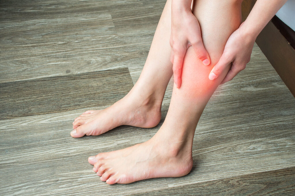
In the earliest stage, changes can be subtle enough to miss, especially on darker skin tones. Look for a faint bluish or slightly darker tint, most often starting around the ankle or lower calf. Mild swelling and a feeling of tightness or heaviness may appear before any obvious color change does.
STAGE 2 · DEVELOPING
As the clot grows, the affected area typically becomes more clearly red, purple, or dusky, with visible swelling that makes one leg noticeably larger than the other. The skin often feels warm to the touch and may look slightly shiny or stretched. Nearby surface veins can appear more prominent than usual.
STAGE 3 · ADVANCED
Left untreated, the skin over a long-standing clot can take on a brownish or darkened tone as broken-down blood cells build up under high pressure, a change called hyperpigmentation. Skin may become dry, tight, or flaky, and in severe cases can develop sores. This stage needs prompt medical attention, not home monitoring.
Where Blood Clots Typically Show Up in the Leg
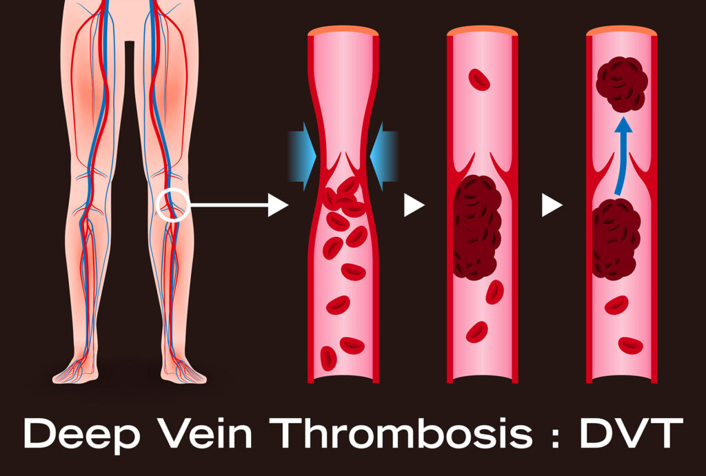
A blood clot in the calf is by far the most common presentation, which is usually what people are picturing when they search for this. Less often, a clot in the leg forms higher up, behind the knee or in the thigh, and tends to be more serious the closer it is to the body.
| Location | How Common | What Makes It Different |
|---|---|---|
| Calf (below the knee) | Most common location | Symptoms can be milder and easier to miss |
| Behind the knee (popliteal) | Common | Swelling often more noticeable, pain with bending |
| Thigh | Less common, more serious | Symptoms tend to be more severe, higher risk of complications |
| Whole leg | Occurs with larger clots | Widespread swelling and color change, higher urgency |
How Doctors Actually Confirm a Blood Clot
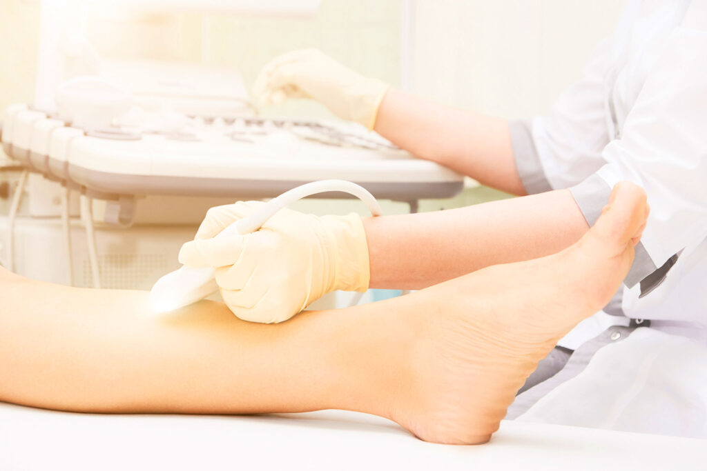
Vein specialists don’t rely on appearance alone; they use a validated scoring tool called the Wells Criteria, first developed in 1995 and refined in the years since. It weighs specific clinical findings, not just how a leg looks, to estimate the probability of DVT before imaging.
| Clinical Factor | Points |
|---|---|
| Active cancer or cancer treatment within 6 months | +1 |
| Paralysis, paresis, or recent leg immobilization | +1 |
| Bedridden 3+ days, or major surgery within 12 weeks | +1 |
| Localized tenderness along the deep venous system | +1 |
| Entire leg swollen | +1 |
| Calf swelling more than 3 cm versus the other leg | +1 |
| Pitting edema confined to the symptomatic leg | +1 |
| Visible collateral superficial veins (non-varicose) | +1 |
| Previously documented DVT | +1 |
| An alternative diagnosis is at least as likely | -2 |
A score of 2 or higher suggests a moderate-to-high probability of DVT and typically leads straight to an ultrasound. A score below 1 makes DVT unlikely, especially when paired with a negative D-dimer blood test. This is exactly why a same-day vein evaluation is more reliable than comparing your leg to a picture online; it accounts for far more than color and swelling alone.
Why a Photo Alone Can’t Diagnose a Blood Clot
Up to half of people with a blood clot show few or no visible signs at all, according to clinical literature indexed by the National Library of Medicine. That means a clot can be present with completely normal-looking skin, and conversely, skin changes that look like a clot can come from a bruise, an infection, or a muscle strain instead. Even a detailed picture of a blood clot in the calf only captures one moment in one person’s case, not the full range of how clots actually look. Appearance is a clue, not a diagnosis.
| Method | What It Can Tell You | Limitation |
|---|---|---|
| Visual inspection or photos | May suggest swelling, color change, or shape irregularities | Can’t detect clots with no visible symptoms; easily confused with bruises or infections |
| Duplex ultrasound | Directly visualizes blood flow and locates the clot | Requires a clinic visit, but is quick, painless, and definitive |
Blood clots in the legs are more common than most people realize. The CDC estimates that as many as 900,000 Americans are affected by venous thromboembolism each year, which is exactly why a systematic evaluation, not a visual guess, is the standard of care.
When Should You See a Doctor of Veins About Blood Clot Symptoms?
See a doctor of veins the same day you notice new swelling, warmth, or color change in one leg, especially if it’s paired with pain that doesn’t ease with rest. A vein specialist can order a duplex ultrasound immediately, which settles the question far faster and more reliably than watching and waiting.
Frequently Asked Questions
What are the first signs of a leg blood clot?
Early on, a faint bluish or reddish tint, mild swelling, and a feeling of heaviness or tightness in the calf are the most common first signs, often before pain sets in.
Can a leg blood clot clear on its own?
Small clots occasionally dissolve naturally, but this isn’t something to rely on. Without treatment, a clot can grow or travel to the lungs, so medical evaluation is always the safer path.
Is it safe to walk with a blood clot in the leg?
Not until a doctor confirms what you’re dealing with. Movement can occasionally help smaller clots resolve, but it can also dislodge a larger one, so this decision should be made with medical guidance, not guesswork.
What does an early stage blood clot look like?
Often subtle: a faint bluish or slightly darker patch of skin, mild swelling, and warmth, most commonly starting around the ankle or lower calf, sometimes without any visible change at all.
A leg that suddenly looks or feels different deserves a same-day answer, not a search engine. Call CURA Vein Doctors at +1 973-363-2029 or reach out through the contact page to get a clear picture, literally.
References
- Waheed SM, Kudaravalli P, Hotwagner DT. Deep Venous Thrombosis. StatPearls [Internet]. StatPearls Publishing, National Library of Medicine. ncbi.nlm.nih.gov/books/NBK507708
- Wells Score for Deep Vein Thrombosis, clinical prediction rule overview. sciencedirect.com/topics/medicine-and-dentistry/wells-score
- Centers for Disease Control and Prevention. Data and Statistics on Venous Thromboembolism. cdc.gov/blood-clots/data-research
- Cleveland Clinic. Deep Vein Thrombosis (DVT): Symptoms, Causes and Treatment. my.clevelandclinic.org/health/diseases/16911-deep-vein-thrombosis-dvt