
Reviewed by Dr. Amir Issa, MD, DABVLM, RPhS · CURA Vein Doctors · October 2026
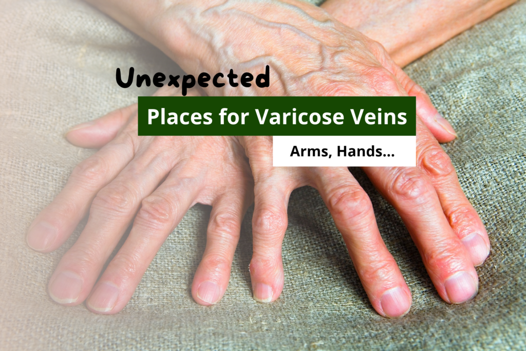
Your legs look fine. So why are the veins on your arms suddenly standing out? Varicose veins have a reputation as a leg problem, but the venous system spans the entire body. When valves fail, or pressure builds, blood pools wherever the anatomy allows. Arms, hands, the face, and the abdomen can all develop visible, dilated veins. Each site has a different cause, a different appearance, and a different set of warning signs.
Which type of prominent vein warrants more than a second glance?
What Causes Veins to Pop Out in Arms and Hands?
1. Varicose Veins on the Hands
Usually benign. Ageing skin, low body fat, heat, and dehydration all make hand veins stand out. Quick test: raise your arm above your head. Normal veins collapse within seconds. Veins that stay prominent, ache, or feel hard deserve a closer look.
True hand varicose veins typically follow prior trauma, repeated IV access, or obstruction further up the arm. Signs to watch: persistent ropiness at rest, skin discoloration over the vein, or a tender, hardened cord along the vein’s path.
2. Varicose Veins on the Arms
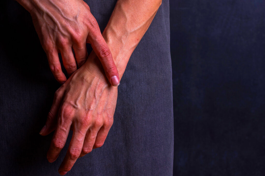
Athletic individuals often develop highly visible arm veins from low body fat and increased blood flow during training. This is vascularity, not disease. Those veins are healthy, functional, and do not need treatment. Purple or blue-tinted veins seen through pale or thin skin are almost always normal superficial veins close to the surface.
The picture shifts when arm veins are persistently dilated, visibly tortuous, or tender in someone who is not heavily muscled. Causes include post-thrombotic vein wall damage, compression of the subclavian vein at the thoracic outlet, and prior catheter or IV line injury. These are not cosmetic findings. A duplex ultrasound is needed to distinguish normal vascularity from genuine reflux or obstruction.
One specific condition that non-specialists often miss: Paget-Schroetter syndrome, also called effort thrombosis. The subclavian vein clots due to repetitive overhead movement in athletes, swimmers, or manual workers. It presents with sudden arm swelling, heaviness, and a bluish discoloration developing over hours. This is a vascular emergency. It is not a finding to monitor or follow up at convenience.3
3. Thread Veins on the Face and Neck
What most people call varicose veins on the face are not varicose veins in the clinical sense. They are telangiectasias, also called thread veins or spider veins: tiny dilated capillaries under 1 mm across, sitting just below the skin surface. True varicose veins are dilated, refluxing vessels above 3 mm. They do not typically occur on the face, and conflating the two leads to the wrong treatment expectation.
Facial telangiectasias are driven by genetics, cumulative sun exposure, rosacea, alcohol use, and hormonal shifts. Valve failure plays no role. The vessels simply dilate and lose the ability to contract fully. Treatment is cosmetic and effective: Nd:YAG laser and intense pulsed light (IPL) are standard approaches. No compression stockings, no phlebectomy, no vein clinic referral in the leg-disease sense. Sun protection and minimizing known triggers slow the rate of new formation.
4. Varicose Veins in the Abdomen and Pelvis
Visible abdominal and groin veins have two very different meanings depending on the pattern. Getting this distinction right matters more than it does anywhere else on this list.
Pelvic congestion syndrome (PCS) affects women, typically after one or more pregnancies. The pelvic veins dilate under sustained hormonal and mechanical load during pregnancy, then fail to recover once pressure is removed. Symptoms include a dull, chronic pelvic ache that worsens with prolonged standing, during menstruation, or after intercourse. It is frequently underdiagnosed because the diseased veins lie deep in the pelvis and cannot be seen externally. Duplex ultrasound or MRI venography confirms it. Transcatheter embolization is the established treatment and avoids the need for open surgery.2
Caput medusae is a different and more serious finding: a radiating pattern of dilated veins spreading outward from the navel. It results from portal hypertension, where liver disease blocks normal portal venous flow, and blood reroutes through collateral abdominal vessels. This is a clinical sign of serious underlying liver pathology. It is not a vein clinic presentation; it needs urgent medical evaluation to identify and manage the cause.
Groin varicosities that persist after pregnancy or worsen with pain and swelling need a duplex assessment. Untreated, a dilated vein under ongoing pressure can rupture or clot; what happens when a vein ruptures explains exactly why early evaluation matters.1
| Location | What you typically see | Common cause | Usually harmless? |
|---|---|---|---|
| Hands | Raised, ropy veins; flattens with arm raised | Ageing, low body fat, heat, dehydration | Usually yes |
| Upper arms | Visible blue-purple veins under skin surface | Vascularity, thin skin, prior IV trauma | Usually yes |
| Face and neck | Fine red or purple web-like vessels | Genetics, sun exposure, rosacea, alcohol | Yes (telangiectasias, not varicose) |
| Lower abdomen and groin | Dilated, tortuous veins; groin bulge | Pelvic congestion syndrome, pregnancy | Needs assessment |
| Around the navel | Radiating vein pattern from navel outward | Portal hypertension, liver disease | No; urgent evaluation required |
When Visible Veins Become a Warning Sign
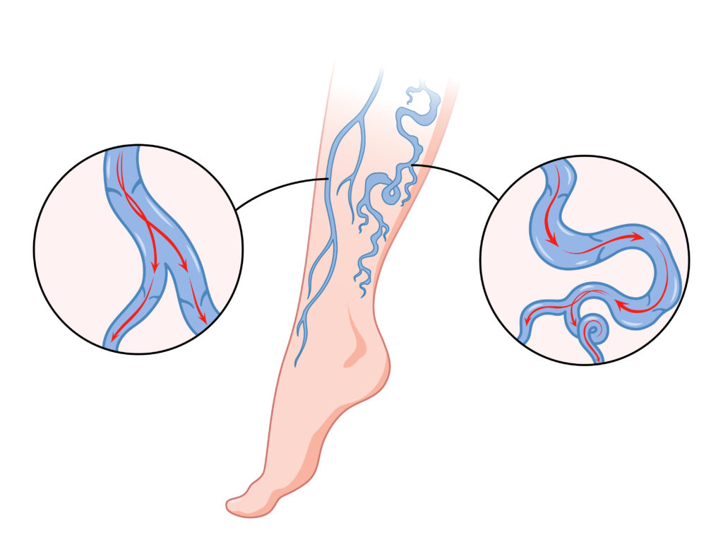
Most visible veins outside the legs are benign. A few patterns indicate something that needs prompt clinical attention. The distinction almost always comes down to the symptoms accompanying the appearance, not the appearance itself. The same venous pressure behind unexplained leg aching can accumulate at unusual sites and drive changes in the arms, abdomen, and face.
| Location | First step | Procedural option |
|---|---|---|
| Hands | Hydration; review heat exposure and grip intensity | Sclerotherapy or phlebectomy for symptomatic veins |
| Arms | Duplex ultrasound to confirm or rule out reflux or obstruction | Foam sclerotherapy; anticoagulation if thrombosis confirmed |
| Face | SPF protection; reduce alcohol and heat triggers | Nd:YAG laser, IPL, or fine-needle sclerotherapy |
| Abdomen and pelvis | MRI venography or duplex ultrasound to map the source | Transcatheter embolization for pelvic congestion syndrome |
What to Expect at a CURA Assessment
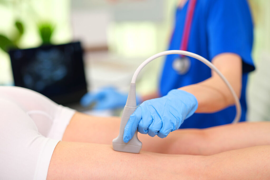
Prominent veins in unusual locations cannot be assessed accurately by appearance. Duplex ultrasound maps the direction of blood flow and identifies valve failure even in veins that look ordinary on the surface. For veins that qualify after ultrasound, sclerotherapy is a safe and effective option across multiple sites, including the hands, arms, and face.
Your Assessment Pathway
Notice prominent or ropy veins
Note whether they are new, persistent, or triggered only by heat and activity.
Elevation test
Raise your arm above your head. Normal superficial veins collapse within seconds. Veins that stay prominent suggest reflux or obstruction.
Check for accompanying symptoms
Aching, swelling, skin changes, or discoloration alongside the visible vein shifts the picture from cosmetic to clinical.
Book a duplex ultrasound
The only accurate way to confirm valve failure, reflux, or obstruction. Appearance alone cannot diagnose vein disease at any site.
Frequently Asked Questions
Why am I all of a sudden getting varicose veins?
A sudden increase usually follows a trigger: pregnancy, significant weight change, prolonged standing, hormonal contraceptives, or a thrombotic event that damaged a valve. Veins that were borderline for years can cross the visible threshold after any of these changes in load or hormonal environment.
Can varicose veins be a symptom of something else?
Yes. Prominent abdominal veins can signal portal hypertension from liver disease. Sudden one-sided arm swelling points to deep vein thrombosis. Pelvic varicosities in women may reflect pelvic congestion syndrome. A duplex ultrasound distinguishes a primary venous problem from one that is secondary to a deeper cause, and if the main complaint is leg pain, it also helps rule out other sources.
What are varicose veins a warning for?
In the legs, varicose veins signal early chronic venous insufficiency, which can progress to skin changes, eczema, and venous ulceration if left untreated. In unusual locations, the concern shifts: arm veins may signal obstruction, and abdominal veins radiating from the navel signal liver pathology requiring urgent medical assessment.
Do varicose veins indicate heart problems?
Not directly. Varicose veins are a peripheral venous disease, not a cardiac or arterial one, and they do not cause heart attacks or strokes. New, rapidly progressive bilateral leg swelling, however, can sometimes overlap with right heart failure and should be evaluated to rule out a cardiac contribution alongside venous disease.
Noticed a vein change in an unexpected place?
A 20-minute duplex ultrasound at CURA Vein Doctors can confirm whether valve failure, reflux, or obstruction is driving what you see. Same-day appointments available in New Jersey.
References
- Eklöf B, Rutherford RB, Bergan JJ, et al. Revision of the CEAP classification for chronic venous disorders: Consensus statement. J Vasc Surg. 2004;40(6):1248–1252.
- Durham JD, Machan L. Pelvic congestion syndrome. Semin Intervent Radiol. 2013;30(4):372–380.
- Illig KA, Doyle AJ. A comprehensive review of Paget-Schroetter syndrome. J Vasc Surg. 2010;51(6):1538–1547.