
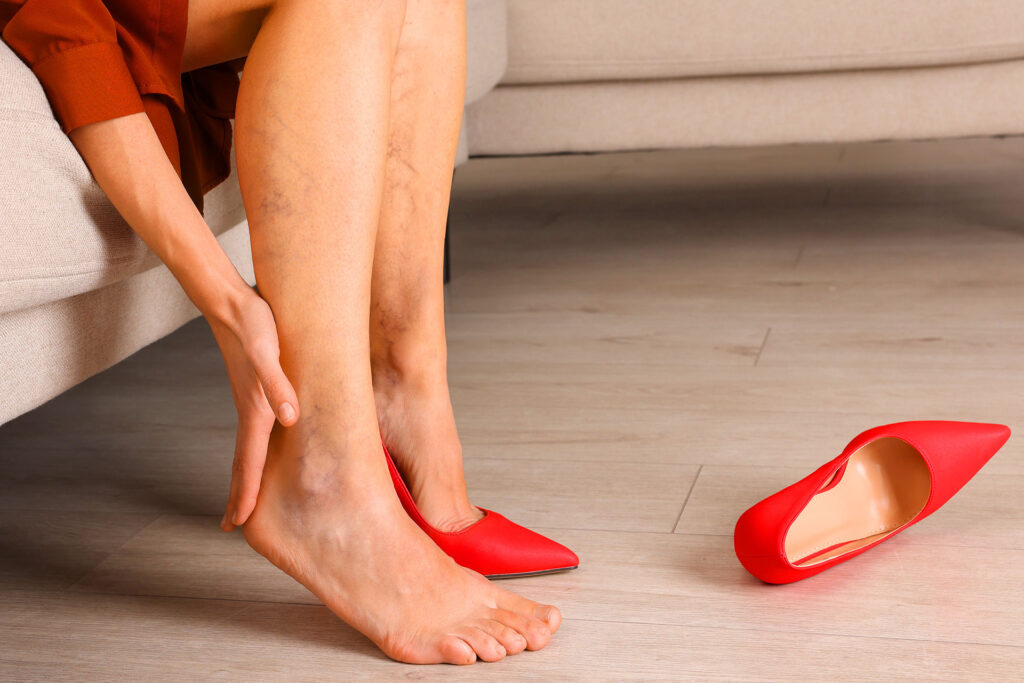
Most people who come in with a burst vein in the leg have been watching it for days. They are not sure if it is serious. They do not know what a specialist actually looks for. And they are surprised when the answer is not just “the bruise.” A burst vein leaves visible damage on the surface. But what drives the rupture is almost always something deeper. Here is what vascular doctors actually look at when a patient walks in with a burst vein in the leg.
What Causes Burst Veins in the Leg?
Burst veins in the leg occur when a vein wall weakens and gives way under pressure, releasing blood into surrounding tissue. The most common driver is chronic venous insufficiency, where failing valves allow blood to pool and press against the vessel wall over time. Trauma, blood thinners, and age accelerate the process. Spontaneous ruptures without injury almost always signal underlying venous disease.
Warning Sign 1: The Rupture Had No Cause
This is the detail doctors focus on first. A bruise from a bump is one thing. A bruise that appeared without contact is another.
If you are an active runner, you might try to link leg pain to your training; see how to manage running-related knee pain here. However, a spontaneous burst vein, occurring with no fall or impact, points directly to a compromised vein wall. The wall did not fail because of an external force. It failed because it was already too weak to hold.
In clinical practice, a single spontaneous rupture in a patient with known varicose veins is expected. Repeated spontaneous ruptures, or a first one in someone with no prior vein history, both warrant a duplex ultrasound to understand what is happening at the valve level.
Warning Sign 2: One Leg Is Larger Than the Other
Asymmetric swelling alongside a burst vein changes the diagnostic picture entirely. When one leg swells notably more than the other, particularly in the calf or thigh, the concern shifts from a surface vein rupture to a possible deep vein thrombosis.
DVT can cause surface veins to dilate as blood reroutes around a blocked deep vessel. The surface vein then appears burst or newly prominent. Treating only the surface in this scenario misses the actual problem entirely.
Any burst vein in the leg with unilateral swelling gets an ultrasound the same day. No exceptions.
Warning Sign 3: The Skin Around the Vein Is Changing
The skin near the inner ankle is where chronic venous disease shows up first. When a burst vein occurs in this area and the surrounding skin is already darkened, hardened, or itching, the rupture is not an isolated event. It is part of a longer disease process.
Lipodermatosclerosis, the hardening of tissue from chronic venous pressure, means the skin has already sustained significant damage. Wounds in this zone heal slowly. A burst vein here carries a real risk of progressing to a venous ulcer if the underlying venous disease is not treated.
⚠️ A burst vein near darkened or hardened ankle skin is not cosmetic. It signals advanced venous disease. Do not treat the surface wound without investigating the underlying pressure driving it. A vein specialist, not a wound clinic, is the right first call.
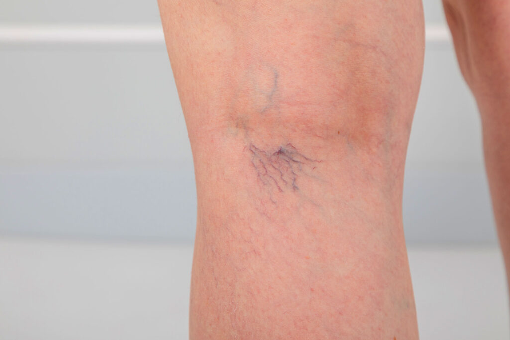
Warning Sign 4: The Vein Feels Like a Cord
After a vein bursts, the body tries to seal it with a clot. Most of the time this is efficient and uneventful. Sometimes the clot triggers inflammation and the vein becomes hard, rope-like, and tender to touch. The skin above it turns red and warm.
This is superficial thrombophlebitis. It is painful, it looks alarming, and it requires treatment with anti-inflammatory medication and compression. It is also a signal that the patient’s venous system is under enough strain to produce clots without significant provocation.
Doctors also check whether the clot has extended into the deep system. Superficial and deep vein involvement can coexist, and the management differs significantly between the two.
Warning Sign 5: It Has Happened Before
A second or third burst vein in the same leg is a pattern. Patterns have causes. In almost every recurring case, the cause is venous insufficiency that has not been addressed.
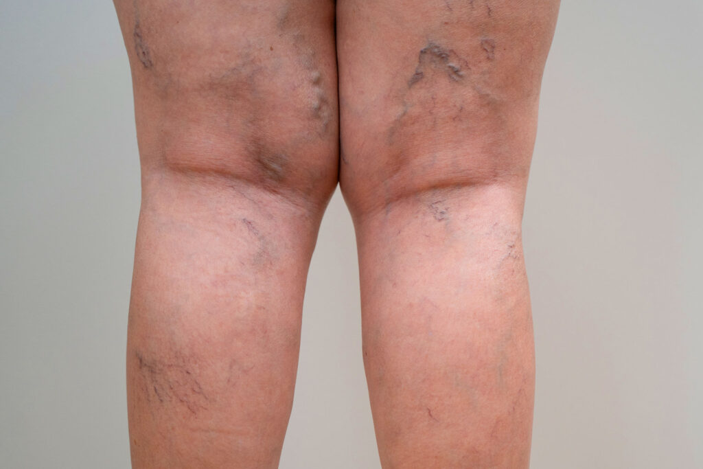
Each rupture causes minor scarring. Each episode of pooled blood leaves hemosiderin deposits in the tissue, the brownish staining visible near the ankle in patients with long-standing venous disease. The damage accumulates quietly until something more serious develops.
Recurring burst veins are one of the clearest clinical indications for vein treatment. Waiting for a more dramatic event is not a medically sound approach.
What the Duplex Ultrasound Actually Shows
Patients often ask why a bruise on the surface requires an ultrasound. The answer is that the bruise is the result. The ultrasound finds the cause.
| What the Ultrasound Checks | What a Normal Result Looks Like | What an Abnormal Result Indicates |
|---|---|---|
| Valve function in the great saphenous vein | Blood flows upward, no reflux | Reflux means CVI is present |
| Deep vein patency | Vein is open, compressible | Non-compressible vein suggests DVT |
| Superficial vein integrity | Smooth, thin walls | Thickened or irregular walls suggest prior damage |
| Flow direction in perforator veins | Inward flow only | Outward flow indicates perforator incompetence |
The results tell the specialist whether the burst vein is a one-off event or the visible result of a failing venous system. The vein treatment plan follows directly from what the imaging shows.
Treatment: Matching the Response to the Cause
| Clinical Finding | Recommended Treatment |
|---|---|
| Minor bruise, no underlying disease | Cold compress, elevation, compression, monitor |
| Superficial thrombophlebitis | Anti-inflammatory medication, compression, ultrasound follow-up |
| Varicose veins driving ruptures | Sclerotherapy, endovenous laser ablation, or radiofrequency ablation |
| CVI confirmed on ultrasound | Ablation of refluxing vein segment, compression therapy |
| DVT confirmed | Anticoagulation, specialist follow-up |
| Skin changes or ulceration present | Vein treatment plus wound care and compression |
✓ Most vein procedures today are outpatient. Endovenous ablation and sclerotherapy take under an hour, require only local anesthesia, and send patients home the same day. The recovery that most people expect simply does not apply to modern vein treatment. If you are struggling with persistent discomfort, it is worth exploring comprehensive leg pain relief strategies that can help you stop the ache and reclaim your stride.
FAQ: Burst Veins
What happens if a vein bursts?
Blood leaks into the surrounding tissue, causing bruising and sometimes a firm swelling called a hematoma. Minor cases resolve in one to two weeks. External bleeding from a varicose vein is less common but more urgent and needs same-day care.
What causes burst veins?
Chronic venous insufficiency and varicose veins are the most common underlying causes. Physical trauma, blood thinners, aging, and clotting disorders all contribute. Spontaneous ruptures without any trauma almost always point to pre-existing venous disease.
What does vein pain feel like?
A dull, heavy ache that builds through the day is the most common description. A burst vein adds sharp tenderness at the rupture site. Thrombophlebitis produces warmth and a cord-like hardness along the vein that is noticeably different from ordinary bruising.
How are spider veins caused?
Spider veins form when small surface vessels dilate under elevated venous pressure. Genetics, hormonal changes, prolonged standing, and valve failure in deeper veins all play a role. In many patients they are a surface signal of a deeper insufficiency driving the pressure from below.
Your Legs Know Something Is Wrong. So Do We.
Burst veins that keep coming back are not bad luck. They are a pattern. At CURA Vein Centers, our specialists find the root cause and treat it directly, not just the bruise it leaves behind.
Free consultation available. No commitment required.