
Reviewed by Dr. Amir Issa, MD, DABVLM, RPhS · CURA Vein Doctors · November 2026
A bruise on your lower leg with no memory of a bump is easy to dismiss the first time. When ecchymosis reappears in the same spots, deepens without a cause, or travels alongside heavy legs and visible veins, something else is driving it. What does your vascular system have to do with it?
What Is Ecchymosis?
Quick Answer
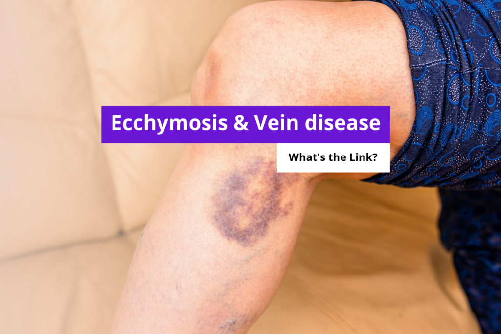
Ecchymosis is a flat, discolored skin patch (purple, red, or brown) formed when blood leaks from ruptured capillaries into surrounding tissue. In vein disease, chronically raised leg pressure weakens capillary walls over time until they rupture without any impact, producing spontaneous bruising on the lower legs.
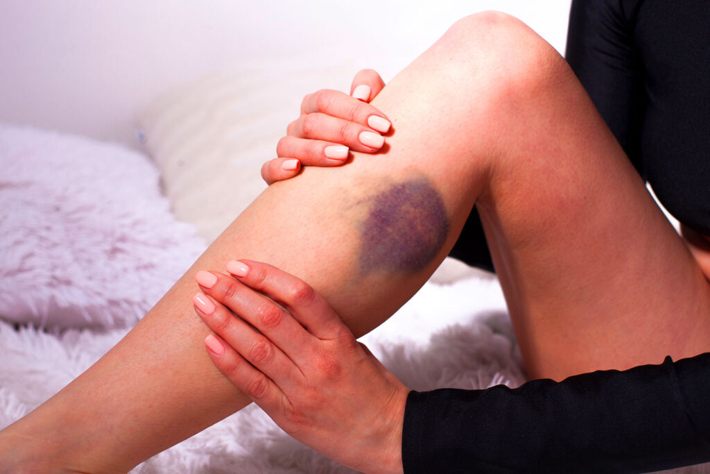
It differs from a hematoma, which pools blood in a raised lump, and from petechiae, which appear as pinpoint dots under 3 mm. Ecchymosis lies flat, spreads over a wider area, and shifts color from deep purple through greenish-yellow as hemoglobin breaks down. That progression happens the same way whether the trigger was a vein or a fall.
How Chronic Venous Insufficiency Causes Ecchymosis in the Legs
Healthy leg veins move blood upward against gravity. When venous valves fail, blood pools in the lower limb and raises hydrostatic pressure inside the microvascular network. That sustained pressure stretches and weakens capillary walls over months and years. Eventually, the walls rupture spontaneously: blood leaks into the dermis and subcutaneous tissue, producing ecchymosis without any external force.[1]
This damage follows a staged progression mapped by the CEAP classification, the global standard for grading chronic venous insufficiency. Spider veins (C1) and varicose veins (C2) are early-stage markers. By C4, sustained venous hypertension has caused visible skin changes including ecchymosis, hyperpigmentation, and dermal thickening. Catching disease before it reaches C4 significantly reduces the risk of permanent skin damage.[1]
| Skin Sign | CEAP Stage | What It Looks Like | What It Signals |
|---|---|---|---|
| Spider veins | C1 | Red or blue threadlike clusters under the skin surface | Early venous reflux; may signal deeper disease beneath |
| Varicose veins | C2 | Bulging, twisted, rope-like veins on the calf or thigh | Moderate valve failure with elevated reflux volume |
| Ecchymosis | C4a–C4b | Flat, irregular purple-brown discoloration with no injury | Elevated pressure breaking down capillary integrity |
| Hemosiderin staining | C4a | Rust-brown patches at the ankle that do not fully fade | Chronic iron deposition from repeated capillary leak |
| Stasis dermatitis | C4a–C4b | Inflamed, scaly, itchy skin over the medial ankle | Prolonged hemosiderin exposure triggering dermal inflammation |
Bruised Vein on Foot and Lower Leg: What the Pattern Tells You
Location and timing narrow the cause. Rust-brown patches clustered around the medial ankle and inner calf, particularly those that do not clear between episodes, usually reflect hemosiderin deposition from long-standing capillary leak.[2] When the same patches become inflamed and scaly, that transition is called varicose eczema and stasis dermatitis, a downstream complication of untreated venous disease.
A vein that looks bruised along its length, appearing darker and discolored without a surface wound, often signals blood pooling within a weakened vessel wall. A bruised vein on the foot that worsens after prolonged standing and improves overnight with elevation points directly to pressure-driven venous stasis. Bilateral involvement, with both legs equally affected, strongly favors a venous or systemic cause over localized trauma.
CVI Skin Progression
Other Causes of Leg Bruising to Rule Out
Vein disease is not the only explanation for easy or spontaneous bruising. Medications are the most common non-vascular trigger. Anticoagulants (warfarin, apixaban, rivaroxaban), daily aspirin, NSAIDs, and long-term oral corticosteroids all impair clotting or thin capillary walls. The bruises they produce can look identical to vein-related ecchymosis. Nutritional deficiencies, particularly vitamins C and K, weaken vessel wall integrity independently of venous pressure. Bleeding disorders such as von Willebrand disease or thrombocytopenia cause bruising patterns that do not follow a gravitational or venous distribution.
| Cause | Typical Pattern | Key Distinguishing Clue | Recommended Next Step |
|---|---|---|---|
| Venous insufficiency | Lower leg and ankle, bilateral, worsens by evening | Visible veins, heaviness, swelling that improves with elevation | Venous duplex ultrasound |
| Anticoagulant or NSAID use | Anywhere on the body, no positional pattern | Correlates with medication start date; no leg-specific symptoms | Review medications with prescriber |
| Vitamin C or K deficiency | Perifollicular petechiae (vitamin C) or large diffuse patches (vitamin K) | No swelling, no visible veins; dietary history is key | Serum vitamin levels; nutritional review |
| Bleeding disorder | Widespread, often arms and torso as well as legs | Family history; prolonged bleeding from minor cuts | CBC, PT/INR, coagulation panel |
| Senile purpura (aging skin) | Forearms and hands most common; thin translucent skin | No swelling or other venous signs; visible skin atrophy | Reassurance; sun protection; monitor for new symptoms |
Spider Vein Removal and Vein Care Options
When ecchymosis links back to venous reflux, treating the reflux source reduces the capillary pressure driving it. Spider vein removal at a vein specialist typically starts with sclerotherapy, a microinjection of sclerosant solution that collapses and seals the damaged vessel. Most patients need one to three sessions spaced four to six weeks apart. For C2 varicose veins feeding the spider network, endovenous thermal ablation addresses the deeper reflux point before surface treatment.[3]
Between appointments, graduated compression (20–30 mmHg) reduces venous hypertension and slows capillary damage. Daily leg elevation above heart level for 20 to 30 minutes complements compression. Ongoing vein care after treatment includes avoiding prolonged standing, maintaining a healthy weight, and wearing compression when traveling. Neither lifestyle measure replaces definitive treatment, but both reduce symptoms and protect skin integrity while awaiting a procedure. Leg aching or heaviness that persists beyond conservative care may reflect a chronic pain pattern worth addressing alongside vein treatment.
Frequently Asked Questions
Is there an autoimmune disease that causes spider veins?
Lupus (SLE), scleroderma, and dermatomyositis all damage small blood vessels and are associated with telangiectasia and spider vein formation. Vasculitis can directly inflame vessel walls, producing visible spider-like patterns alongside joint pain, rash, or other systemic symptoms that warrant rheumatology evaluation alongside vein care.
What health conditions cause spider veins?
Chronic venous insufficiency is the most common cause, but thyroid disease, rosacea, liver cirrhosis, connective tissue disorders, and prolonged corticosteroid use all increase spider vein formation. Hormonal shifts from pregnancy, oral contraceptives, and estrogen therapy significantly raise the risk as well.
What vitamin deficiency causes spider veins?
Vitamin C deficiency weakens collagen in vessel walls, making capillaries fragile and prone to visible leakage and spider vein formation. Low vitamin K and B-vitamin deficiencies, particularly folate and B12, are also linked to endothelial dysfunction that promotes telangiectasia over time.
How to get rid of spider telangiectasia?
Sclerotherapy is the most effective treatment for leg telangiectasia, clearing veins in one to three sessions with minimal downtime and a high clearance rate. Laser therapy (Nd:YAG or pulsed dye) works well for facial and very fine vessels where injection needles are impractical.
Ecchymosis on Your Legs? Get a Vein Evaluation.
Spontaneous bruising on the lower legs is a known sign of advancing venous disease. A duplex ultrasound takes under an hour and tells you exactly where the reflux is.
References
- Lurie F, Passman M, Meisner M, et al. The 2020 update of the CEAP classification system and reporting standards. J Vasc Surg Venous Lymphat Disord. 2020;8(3):342–352. doi:10.1016/j.jvsv.2019.12.075
- Rabe E, Guex JJ, Puskas A, et al. Epidemiology of chronic venous disorders in geographically diverse populations: results from the Vein Consult Program. Int Angiol. 2012;31(2):105–115.
- Khilnani NM, Grassi CJ, Kundu S, et al. Multi-society consensus quality improvement guidelines for the treatment of lower-extremity superficial venous insufficiency with endovenous thermal ablation. J Vasc Interv Radiol. 2010;21(1):14–31. doi:10.1016/j.jvir.2009.10.022